Svc Syndrome Physical Exam
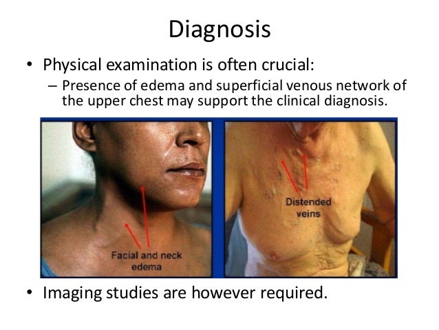
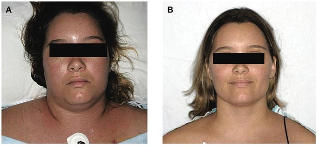
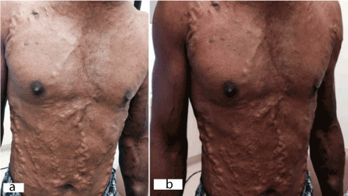
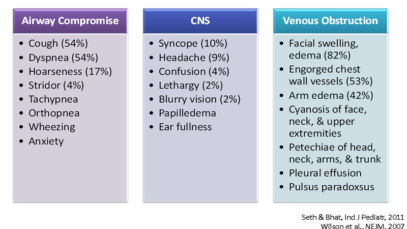
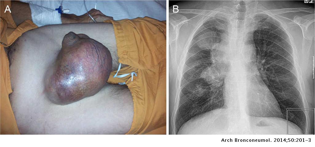
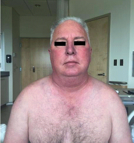
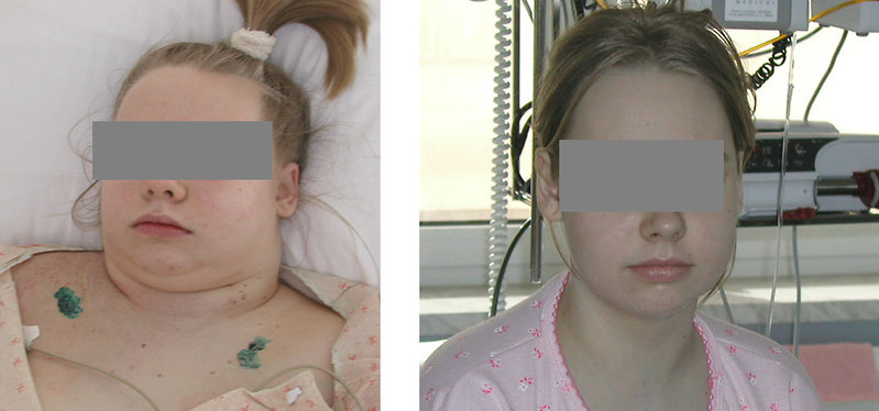
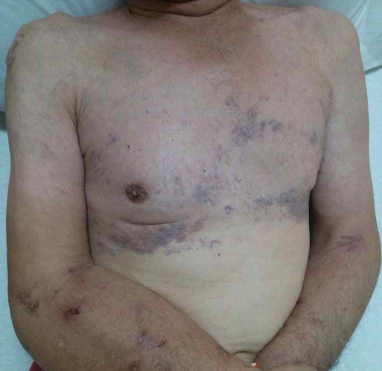
Svc syndrome physical exam. 1 The patient suffered from metastatic renal cell carcinoma which is a rare and atypical cause of SVC syndrome. The most common presenting symptoms of SVC syndrome are faceneck swelling distended neck veins cough dyspnea orthopnea upper extremity swelling distended chest vein collaterals and conjunctival suffusion. Facial neck and even arm swelling.
It carries blood from your head neck upper chest and arms to the heart. For example symptoms such as getting up in the morning with a swollen face and neck edemaswelling without other explanations are important clues. Superior vena cava syndrome SVCS is defined by the obstruction of blood flow through the superior vena cava SVC as a result of intraluminal.
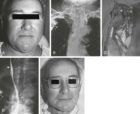
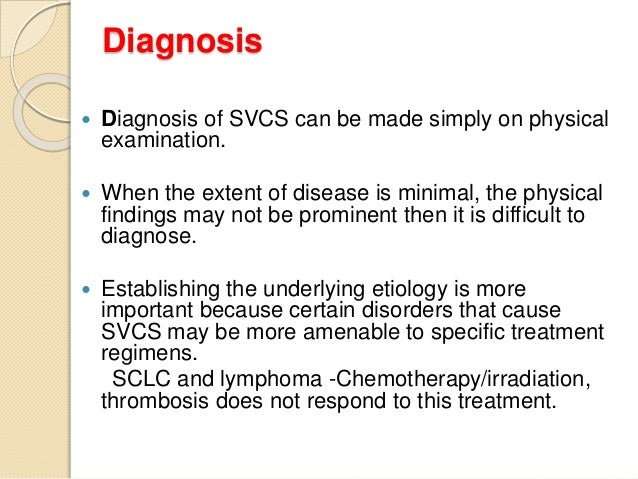
If a doctor suspects a person has superior vena cava syndrome they will first do a physical exam. A careful physical examination is often sufficient to rule out a cardiogenic origin to the patients symptoms. The main techniques of diagnosing SVCS are with chest X-rays CXR CT scans transbronchial needle aspiration at bronchoscopy and mediastinoscopy.
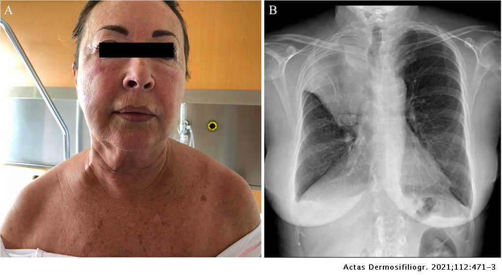
BILYEUSUPERIOR VENA CAVA SYNDROME 351 as in our discussion case any of the following list of symptoms may ensue. Chest radiograph of a 50-year-old woman with complaint of shortness of breath and facial. Superior vena cava SVC syndrome is the result of stenosis or occlusion of the SVC or bilateral bra-chiocephalic veins.
Other non-malignant causes include thrombus secondary to vascular catheterisation goitre or mediastinal fibrosis. A CT image showing compression of the right hilar structures by cancer. Chest radiograph of a patient with known superior vena cava syndrome SVCS and bronchogenic carcinoma CA.
The clinical diagnosis of SVC syndrome is based largely on history and physical examination. And in severe cases seizures syncope or even coma. In a recent series review the average time from the onset of symptoms of SVC ob-.
The exam may show enlarged veins in the upper body. The symptoms that result from compression of the superior vena cava are known as superior vena cava syndrome.
Adenocarcinoma of the lung is the most common cause.
The superior vena cava is a major vein in your upper body. A CT image showing compression of the right hilar structures by cancer. Superior vena cava syndrome SVCS happens when the superior vena cava is partially blocked or compressed. It carries blood from your head neck upper chest and arms to the heart. The clinical diagnosis of SVC syndrome is based largely on history and physical examination. The drawing below shows where the superior vena cava is in your body. The diagnosis of SVC syndrome is usually clinical and requires a high degree of suspicion. Symptoms and patient history are likely the most important ways to diagnose the disease. Adenocarcinoma of the lung is the most common cause.
And in severe cases seizures syncope or even coma. The drawing below shows where the superior vena cava is in your body. The diagnosis of SVC syndrome is usually clinical and requires a high degree of suspicion. Superior vena cava SVC syndrome is the result of stenosis or occlusion of the SVC or bilateral bra-chiocephalic veins. A CXR of a person with lung cancer which was causing superior vena cava syndrome. It carries blood from your head neck upper chest and arms to the heart. Currently the origin is generally cancer or thrombotic events.


















Posting Komentar untuk "Svc Syndrome Physical Exam"